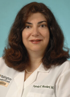
Pamela Woodard, MD

Published June 22, 2020
Pamela Woodard, MD, is a professor of radiology and biomedical engineering and senior vice chair and division director of Radiology Research Facilities. Her areas of specialty include cardiac CT and MRI, and pulmonary embolism diagnosis.
Dr. Woodard sees patients at:
- Mallinckrodt Institute of Radiology, 510 S. Kingshighway
Please call 314-362-7111 to schedule an appointment.
What happened in the course of schooling to make you choose your specialty – cardiothoracic radiology?
During my medical school rotations at Duke University, I had an attending physician who was board-certified in radiology and medicine. He actively practiced both and this was very unusual. I was fascinated with the way he could read an image to provide answers that weren’t obvious in the physical exam.
I always thought I would go into some type of internal medicine subspecialty, but this doctor impressed me with what radiology brought to the clinical table and how imaging results had a direct impact on patient care.
Duke also required a year of research as part of the four-year medical school program. The first year of school was basic science courses, the second year was clinical rotations, the third year was research, and the fourth year was a return to clinical rotations.
My year of research was a turning point – it gave me exposure to basic concepts in grant writing and research study design. I decided I probably wasn’t going to be a basic science researcher, but I did like translational research – applying what I learned in the lab directly to patient care.
What brought you to Washington University?
I came here in 1995 as a cardiothoracic fellow in radiology at the Mallinckrodt Institute of Radiology. At the end of my fellowship, I stayed on as faculty.
Can you explain the role of a diagnostic radiologist?
A diagnostic radiologist uses imaging to look within the body for abnormalities in specific organs. In my job in cardiac imaging, I use cardiac magnetic resonance imaging (MRI) and computed tomography (CT) imaging to look at the anatomy and diseases of the heart. These tests might be performed to look for coronary artery disease in certain patients, or to further assess an abnormality that the cardiologist found on physical examination or on another test.
Which aspect of your practice is most interesting?
Cardiac CT and MR imaging are fascinating. Often cardiac CT and MRI are requested when there is a diagnostic dilemma that can’t be answered by a physical examination or other testing. Our team sees some of the most challenging cardiac imaging cases. It’s very rewarding to help a patient with a correct diagnosis.
Can you explain why it is so important to have a specialist read the CT or MRI?
I don’t think patients realize that there are differences in the quality of the interpretation. One of the benefits of having your CT or MRI performed at Barnes-Jewish hospital by a Washington University radiologist is that it is read by someone who is a specialist in his or her particular modality or organ system. So the radiologist who looks at your cardiac MRI is reading cardiac MRIs the majority of his or her time – not just occasionally.
Having a specialist interpret the study is to the advantage of the patient. Because of our size and experience, there are conditions we see many times and are familiar with, compared to a smaller practice that might only see a particular condition once or twice in the life of their practice. You certainly don’t want to be the first case with a condition that a less experienced radiologist has to diagnose.
What new developments in your field are you most excited about?
We have just purchased a combined PET-MR imaging tool that allows for simultaneous collection of PET and MRI data. We hope that the PET-MRI, right now one of only four in the United States, will be a platform for translational research and imaging for our doctors and patients.
The biggest advantage of the PET-MRI is it doesn’t give ionizing radiation. The biggest use will be for oncologic (cancer) imaging because of the decreased radiation dose – especially important for pediatric patients.
Did you always know you wanted to be a doctor?
I wanted to be a physician ever since I was three or four years old – when my brother was born. I was fascinated to know there were doctors who delivered babies. But I wasn’t sure I wanted to stay up all night, every night.
There were several woman physicians who were influential when I was a child. My mother’s obstetrician was a woman and there was a woman surgeon at the hospital where my dad was an administrator.
The surgeon’s name was Tenley Albright, MD – she was the one of the first women to attend Harvard Medical School. Before Harvard, she won the gold medal for figure skating in the 1956 Winter Olympics — the first American female skater to do so.
Dr. Albright was a resident in the hospital where I was born and my parents have a picture of her holding me as a baby.
Is there an award or achievement that is most gratifying?
My appointment as director for the Center for Clinical Imaging Research (CCIR) is very exciting. Some of the most advanced imaging and translational research takes place in the CCIR.
There is an article that just came out in the New England Journal of Medicine about a clinical study we did. Washington University was one of several sites that studied acute chest pain in the emergency room. We demonstrated that patients who had a coronary computed tomography angiogram (CCTA), as opposed to the routine standard of care, were discharged from the emergency room faster.
I’ve also been a Best Doctor every year since 2005 – that’s very gratifying.
Where are you from?
I grew up in Newton, Massachusetts, then lived in North Carolina for 13 years during college, medical school, internship and residency.
If you weren’t a doctor, what would you be doing?
Technically that means I would be practicing medicine without a license . . . or I’d be a foreign news correspondent for CNN.